5.8 Virtual Gel
The Virtual Gel tab will appear above the sequence viewer when nucleotide documents containing fewer than 50 sequences or 1000 restriction sites are selected.
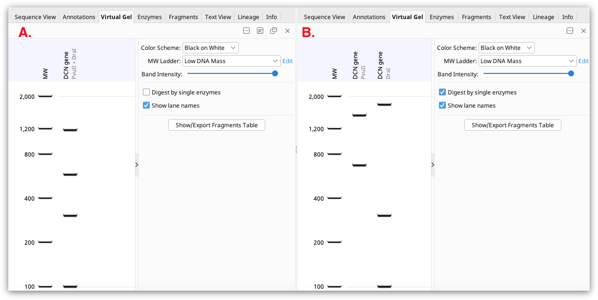
If a single sequence annotated with restriction enzyme sites is selected, the gel displays the fragments that would result from that restriction digest. If the restriction sites are all from a single enzyme, the digest pattern is shown in one lane of the gel. If multiple different restriction enzymes are annotated, the results of digestion with each enzyme will be shown in different lanes if Digest by single enzymes is checked. If this option is unchecked, the result will be shown as a multiple enzyme digest in a single lane (see Figure 5.8
).
If multiple sequence documents containing restriction sites are selected (either by bulk-selecting individual sequence documents, or selecting a sequence list), digests for each sequence are shown in separate lanes on the gel (a maximum of 50 sequences can be shown).
The fragment sizes of all bands shown on the gel can be viewed in the Fragments table or by clicking Show/Export Fragments table. This table is only available for sequences with annotated restriction sites.
For sequences which do not have annotated restriction sites, these will be shown in individual lanes on the gel if the sequences are in separate documents, or in one lane of the gel if the sequences are contained in a list (for example the results of the Digest into Fragments operation).
By default, sequences will appear on the gel in the order they are displayed in the document table or sequence list. To change the order of display, simply drag and drop the sequences on the gel to the desired position.
Note that if uncut circular DNA is selected (for example a plasmid sequence with no restriction sites), the band on the gel will show the size of the linear fragment, rather than the supercoiled DNA size. Depending on the size of your DNA and the buffer you use, uncut circular DNA may migrate differently to linear DNA.